Triangular Fibrocartilage Complex Injury Treatment
The triangular fibrocartilage complex (TFCC) is a structure made up of cartilage and ligaments located on the pinky side of the wrist. The TFCC stabilizes, supports and cushions the wrist’s carpal bones so that you can rotate your arm or grasp objects. When you experience an injury or tear in this region, chronic pain is a common symptom.
At Hand Center of Louisiana, patients in New Orleans and the Gulf South who suspect that they may have a TFCC tear can have a cone beam CT arthrogram performed conveniently in-house. This imaging technique is a far more comfortable and cost-effective alternative to MRI while still allowing your doctor to spot minute injuries to the TFCC.

What Causes Triangular Fibrocartilage Complex Injuries?
Anyone can experience an injury to the TFCC. The most common cause is trauma sustained when someone falls and lands on their outstretched hand, thus bending the wrist backward. However, athletes such as gymnasts, who practice excessive wrist rotation, can also be at risk. TFCC injuries may also be degenerative, occurring over time with age. Inflammatory disorders, like rheumatoid arthritis, can contribute to TFCC tears as well.
New Procedure for Advanced Diagnosis of TFCC
TFCC tears can be tough to diagnose. However, at Hand Center of Louisiana, we use an advanced imaging procedure called cone beam CT arthrogram to examine the thin structures of the triangular fibrocartilage complex.
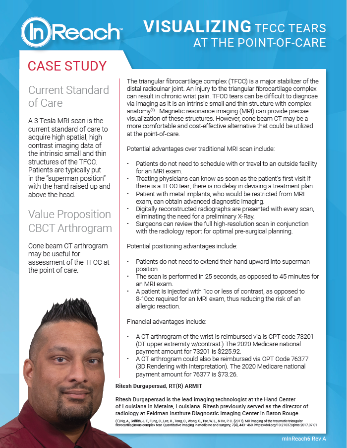
The current standard of care involves a preliminary X-ray to rule out fractures and other abnormalities. Then, magnetic resonance imaging (MRI) provides precise visualization of the minute structures of the TFCC. Unfortunately, to obtain this high-resolution imaging data, patients are placed in the uncomfortable “superman position” for nearly 45 minutes, with their hands raised above their heads.
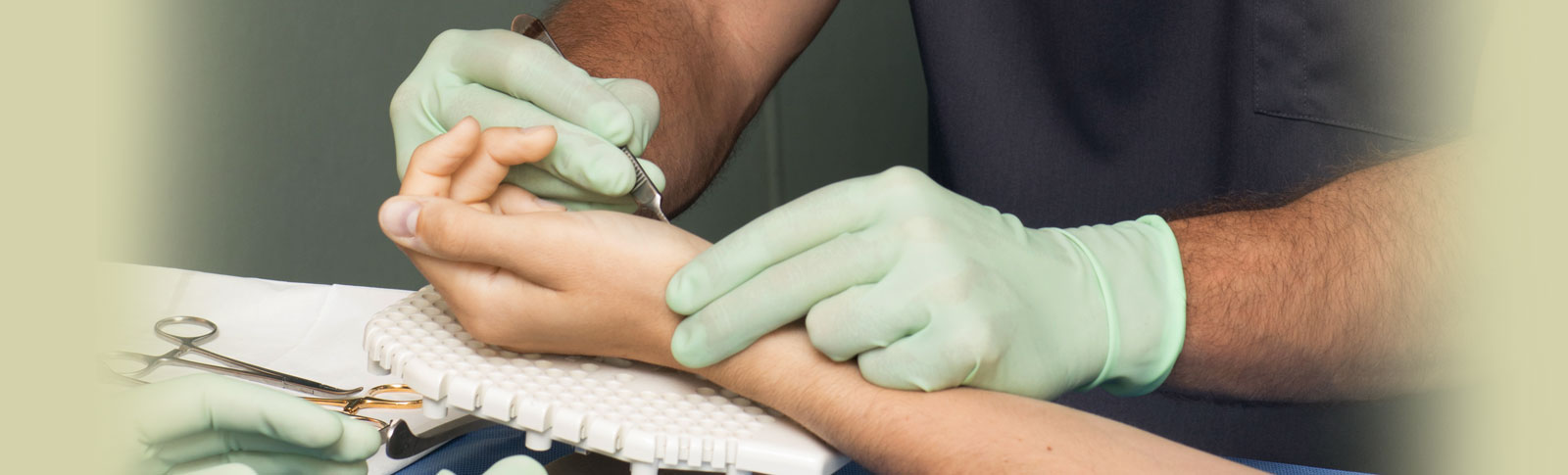
With a cone beam CT arthrogram, patients can be seated comfortably with their wrist placed inside the imaging equipment. The procedure requires 1cc or less of contrast, as opposed to the 8 to 10cc needed for an MRI exam, thus reducing the risk of an allergic reaction. In just 25 seconds, the scan is complete with no recovery time required.
Other significant advantages of getting a cone beam CT arthrogram at Hand Center of Louisiana include the following:
- There’s no need to schedule an MRI exam at an outside facility and no
need for a preliminary X-ray. Your procedure is performed in-house by
our friendly, knowledgeable lead imaging technologist. - Your physician will know right after the procedure whether there is a
TFCC tear and will be able to design your treatment plan as soon as
possible. - Unlike MRI, those with metal implants can still obtain diagnostic
imaging. - The high-resolution scan ensures optimal pre-surgical planning.
TFCC Tear Treatment in New Orleans
The specialists at Hand Center of Louisiana are here to ensure you receive the most advanced treatment options for triangular fibrocartilage complex injuries and tears. Your individual plan may include nonsurgical techniques, such as:
- Splints.
- Medication.
- Injections.
- Physical therapy.
However, if your condition does not respond to these methods, our New Orleans based physicians are highly trained in advanced surgery to repair the TFCC.
Experiencing Chronic Wrist Pain? Schedule Your Appointment at Hand Center of Louisiana
Don’t allow chronic wrist pain to take away from your quality of life and peace of mind. We invite patients throughout New Orleans and the entire Gulf South to schedule an appointment at Hand Center of Louisiana, where you can learn more about cone beam CT arthrogram.